Department of Electrical & Computer Engineering
Signal and Image Laboratory (SaIL)
The University of Arizona®
|
|
|
|
Past Research |
Automated TAPSE Computation in Cardiac MRI
Student: Srinivas L. Naik, Liangchh "James" Huang
| |
Assessing the volume of right ventricle (RV) and its range of movement
during a cardiac cycle currently requires a cardiologist to tediously
track the RV boundary in the cardiac magnetic resonance (CMR) image
sequence throughout a cardiac cycle. Even with simplified measurement
such as tricuspid annulus plane systole excursion (TAPSE),
 manually
identifying associated anatomical landmarks still makes the analysis
procedure inefficient and inconsistent. To automate this procedure, we
need an algorithm to automatically track the change in pixel
coordinates for each such landmark as it moves from one image to the
next during the cardiac cycle. However, existing registration-based
cardiac motion estimation technique, which generally compute the
motion for every pixel in the image, are quite complex and require an
impractical amount of computation.
In order to make this calculation
more feasible for widespread clinical use, our specific aim was to
design a new non-rigid registration algorithm to estimate the motion
of anatomical landmarks with high efficiency and the ability to handle
highly nonlinear motion such as RV motion. This tracking system
enabled us to efficiently explore new cardiac indices which better
correspond to RV function. Finally, by combining the motion estimation
from different image views, we created a linear RV volume
estimator, which is more efficient and consistent than
relying on manually drawn boundaries, and which potentially will have better
accuracy than the inference from the single distance between apex and
tricuspid annulus plane.
In this work, we implemented a preliminary automatic landmark
tracking system and demonstrated it on a long-axis four-chamber-view
CMR image sequence. Motion vectors are estimated for each pixel
between two successive local sub-images. Then, by integrating the
motion vectors through time, we obtain the trajectory of the landmarks
defined in the first image frame.
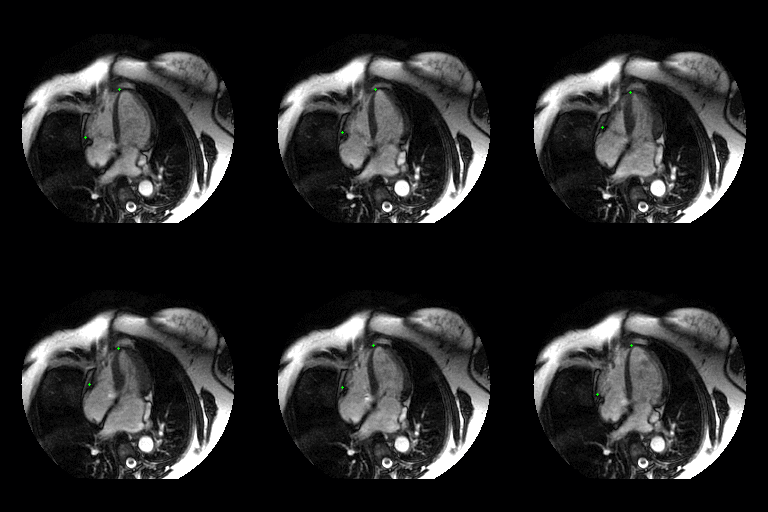
The video shown above is tracking apex (top-right) and the lateral
TA plane (bottom-left) (shown as two small green dots) from end-diastole landmark locations through the entire cardiac cycle.
The image contrast has been enhanced for visual display.
This work was a collaborative effort with Dr. Vincent L. Sorrell in the College of Medicine at The University of Arizona.
Publications:
-
Srinivas L. Naik, Jeffrey J. Rodriguez, Nishant Kalra, and Vincent L. Sorrell, "Tricuspid Annular plane Systolic Excursion (TAPSE) Revisited Using CMR," Journal of Cardiovascular Magnetic Resonance, 2012, vol. 14 (Suppl 1), Feb. 1, 2012, p. 299. doi: 10.1186/1532-429X-14-S1-P299. Presented at the Society for Cardiovascular Magnetic Resonance (SCMR) 15th Annual Scientific Sessions, Orlando, FL, Feb. 2-5, 2012. [PDF ]
-
James L. Huang and Jeffrey J. Rodriguez "Non-Rigid Registration Using Gradient of Self-Similarity Response," Image and Vision Computing, vol. 32, no. 11, Nov 2014, pp. 825-834. [ PDF ]
|
|
1230 E. Speedway Blvd., P.O. Box 210104, Tucson, AZ 85721-0104
|
©2014 All Rights Reserved. |
| Contact webmaster
|
|
|
|
|
