Traumatic brain injury
Low-Rank Representation of Head Impact Kinematics: A Data-Driven Emulator |

|
Summary: Head motion induced by impacts has been deemed as one of the most important measures in brain injury prediction, given that the vast majority of brain injury metrics use head kinematics as input. Recently, researchers have focused on using fast approaches, such as machine learning, to approximate brain deformation in real time for early brain injury diagnosis. However, training such models requires large number of kinematic measurements, and therefore data augmentation is required given the limited on-field measured data available. In this study we present a principal component analysis-based method that emulates an empirical low-rank substitution for head impact kinematics, while requiring low computational cost. In characterizing our existing data set of 537 head impacts, each consisting of 6 degrees of freedom measurements, we found that only a few modes, e.g., 15 in the case of angular velocity, is sufficient for accurate reconstruction of the entire data set. Furthermore, these modes are predominantly low frequency since over 70% of the angular velocity response can be captured by modes that have frequencies under 40 Hz. We compared our proposed method against existing impact parametrization methods and showed significantly better performance in injury prediction using a range of kinematic-based metrics—such as head injury criterion (HIC), rotational injury criterion (RIC), and brain injury metric (BrIC)—and brain tissue deformation-based metrics—such as brain angle metric (BAM), maximum principal strain (MPS), and axonal fiber strains (FS). In all cases, our approach reproduced injury metrics similar to the ground truth measurements with no significant difference, whereas the existing methods obtained significantly different values as well as substantial differences in injury classification sensitivity and specificity. This emulator will enable us to provide the necessary data augmentation to build a head impact kinematic data set of any size. |
Dynamics of human brain and skull |

|
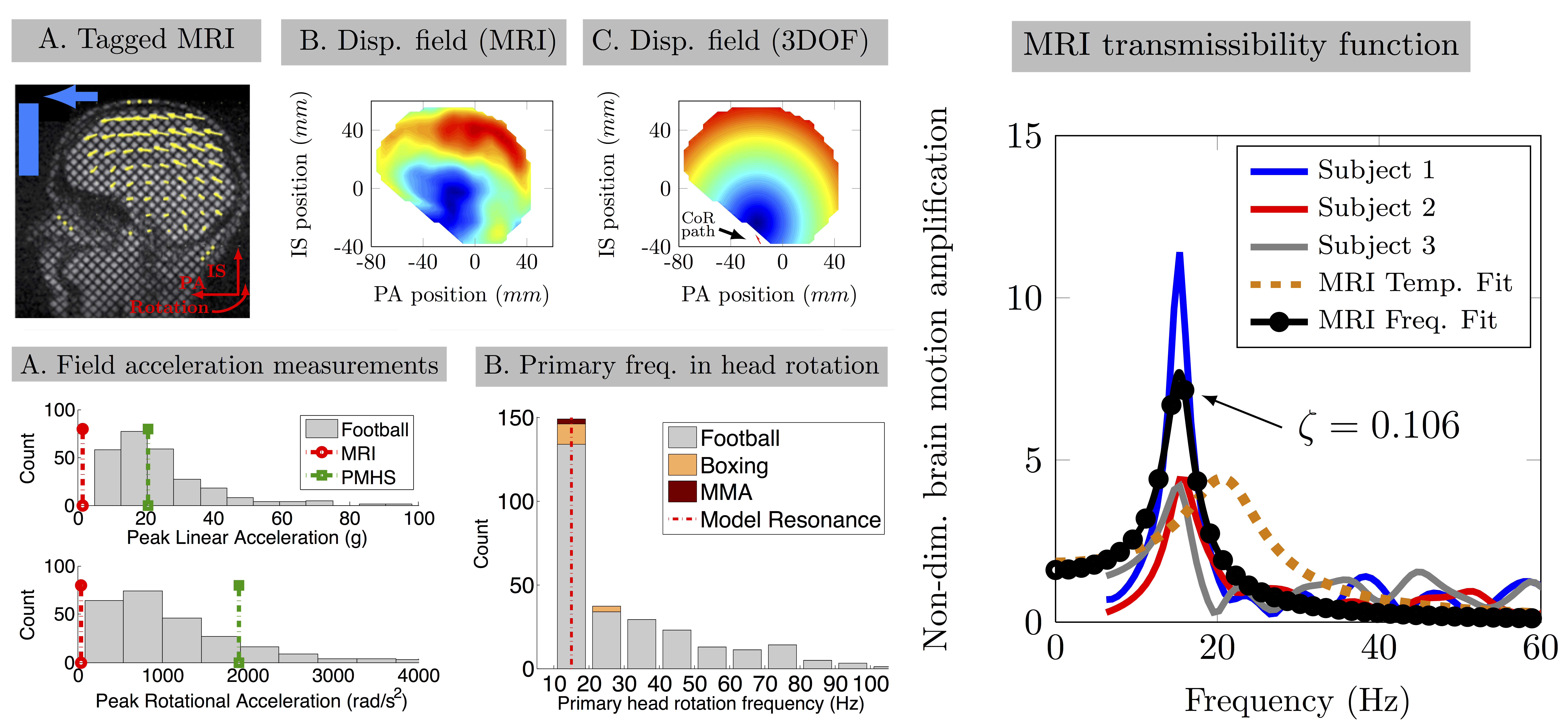
Summary: Although safety standards have reduced fatal head trauma due to single severe head impacts, mild trauma from repeated head exposures may carry risks of long-term chronic changes in the brain's function and structure. To study the physical sensitivities of the brain to mild head impacts, we developed the first dynamic model of the skull–brain based on in vivo MRI data. We showed that the motion of the brain can be described by a rigid-body with constrained kinematics. We further demonstrated that skull–brain dynamics can be approximated by an under-damped system with a low-frequency resonance at around 15 Hz. Furthermore, from our previous field measurements, we found that head motions in a variety of activities, including contact sports, show a primary frequency of less than 20 Hz. This implies that typical head exposures may drive the brain dangerously close to its mechanical resonance and lead to amplified brain–skull relative motions. Our results suggest a possible cause for mild brain trauma, which could occur due to repetitive low-acceleration head oscillations in a variety of recreational and occupational activities. |
Neuroimaging in biomechanics |

|
Summary: We measure and model in vivo brain motion and investigate how we can minimize sports brain trauma. Designing protective devices for the head has long focused primarily on better helmet padding material to prevent skull deformation (occurring at ~100Hz), while overlooking the dynamics of skull-brain. As such, helmets are effective in protecting the skull. However, we have shown that brain’s motion is dominated by slow dynamics (~20Hz) and it can be driven dangerously close to its resonance frequency in typical head impacts in sports such as soccer and football. This observation undermines the effectiveness of current helmets in repeated low-energy head impacts and necessitates systematic investigation of brain motion dynamics. |

|
Summary: The brain’s stiffness measurements from magnetic resonance elastography (MRE) strongly depend on actuation frequencies, which makes cross-study comparisons challenging. We performed a preliminary study to acquire optimal sets of actuation frequencies to accurately obtain rheological parameters for the whole brain (WB), white matter (WM), and gray matter (GM). Six healthy volunteers aged between 26 and 72 years old went through MRE with a modified single-shot spin-echo echo planar imaging pulse sequence embedded with motion encoding gradients on a 3T scanner. Frequency-independent brain material properties and best-fit material model were determined from the frequency-dependent brain tissue response data (20 -80 Hz), by comparing four different linear viscoelastic material models (Maxwell, Kelvin-Voigt, Springpot, and Zener). During the material fitting, spatial averaging of complex shear moduli (G*) obtained under single actuation frequency was performed, and then rheological parameters were acquired. Since clinical scan time is limited, a combination of three actuation frequencies that would provide the most accurate approximation and lowest fitting error was determined for WB, WM, and GM by optimizing for the lowest Bayesian information criterion (BIC). BIC scores for the Zener and Springpot models showed these models approximate the multifrequency response of the tissue best. The best-fit frequency combinations for the reference Zener and Springpot models were identified to be 30-60-70 and 30-40-80 Hz, respectively, for the WB. Optimal sets of actuation frequencies to accurately obtain rheological parameters for WB, WM, and GM were determined from shear moduli measurements obtained via 3-dimensional direct inversion. We believe that our study is a first-step in developing a region-specific multifrequency MRE protocol for the human brain. |
Multi-directional dynamic model for traumatic brain injury detection |

|
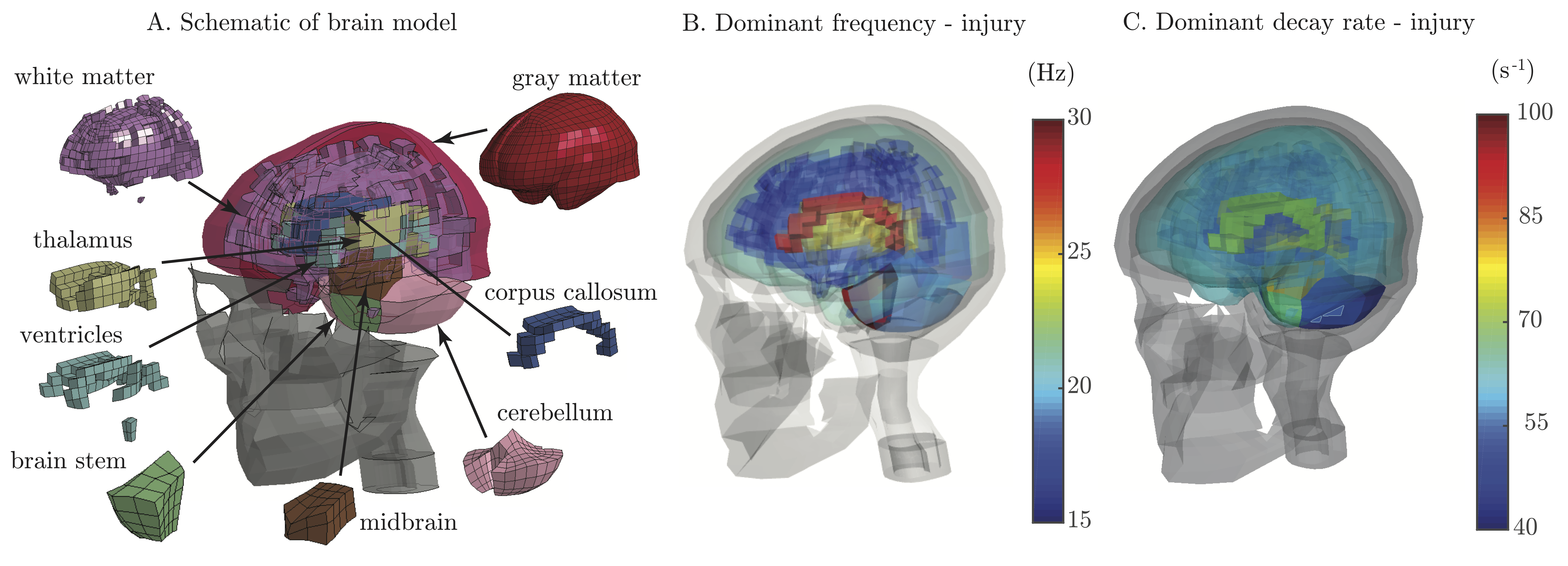
Summary: Given the worldwide adverse impact of traumatic brain injury (TBI) on the human population, its diagnosis and prediction are of utmost importance. Recently, there has been a push toward using computationally expensive finite element (FE) models of the brain to create tissue deformation metrics of brain injury. Here, we develop a new brain injury metric, the brain angle metric (BAM), based on the dynamics of a 3 degree-of-freedom lumped parameter brain model. The brain model is built based on the measured natural frequencies of an FE brain model simulated with live human impact data. We show that it can be used to rapidly estimate peak brain strains experienced during head rotational accelerations that cause mild TBI. In our data set, the simplified model correlates with peak principal FE strain and fiber-oriented peak strain in the corpus callosum on a data set of head kinematics from 27 clinically diagnosed injuries and 887 non-injuries. Our brain model can be used to rapidly approximate the peak strain resulting from mild to moderate head impacts and to quickly assess brain injury risk. |
Measure pathophysiology immediately following head trauma using photoacoustic and ultrasound imaging |

|
Summary: Within minutes following head trauma, a dynamic collection of physiologic and cellular alterations rapidly emerges to initiate a course of pathology and protection that will ultimately define patient outcomes in terms of survival, recovery and long-term disability. Currently, the pre-hospital management of severe TBI – which accounts for 288,000 of the nearly 2.5 million TBI cases annually in the United States and over 57,000 deaths – lacks both diagnostic imaging markers and effective immediate intervention. Thus, rapid, on-site mapping of vulnerable brain tissue, prediction of clinical outcomes close to time of injury (the Golden Hour) can greatly reduce morbidity and mortality following head trauma. |
New perspectives on protective equipment |

|
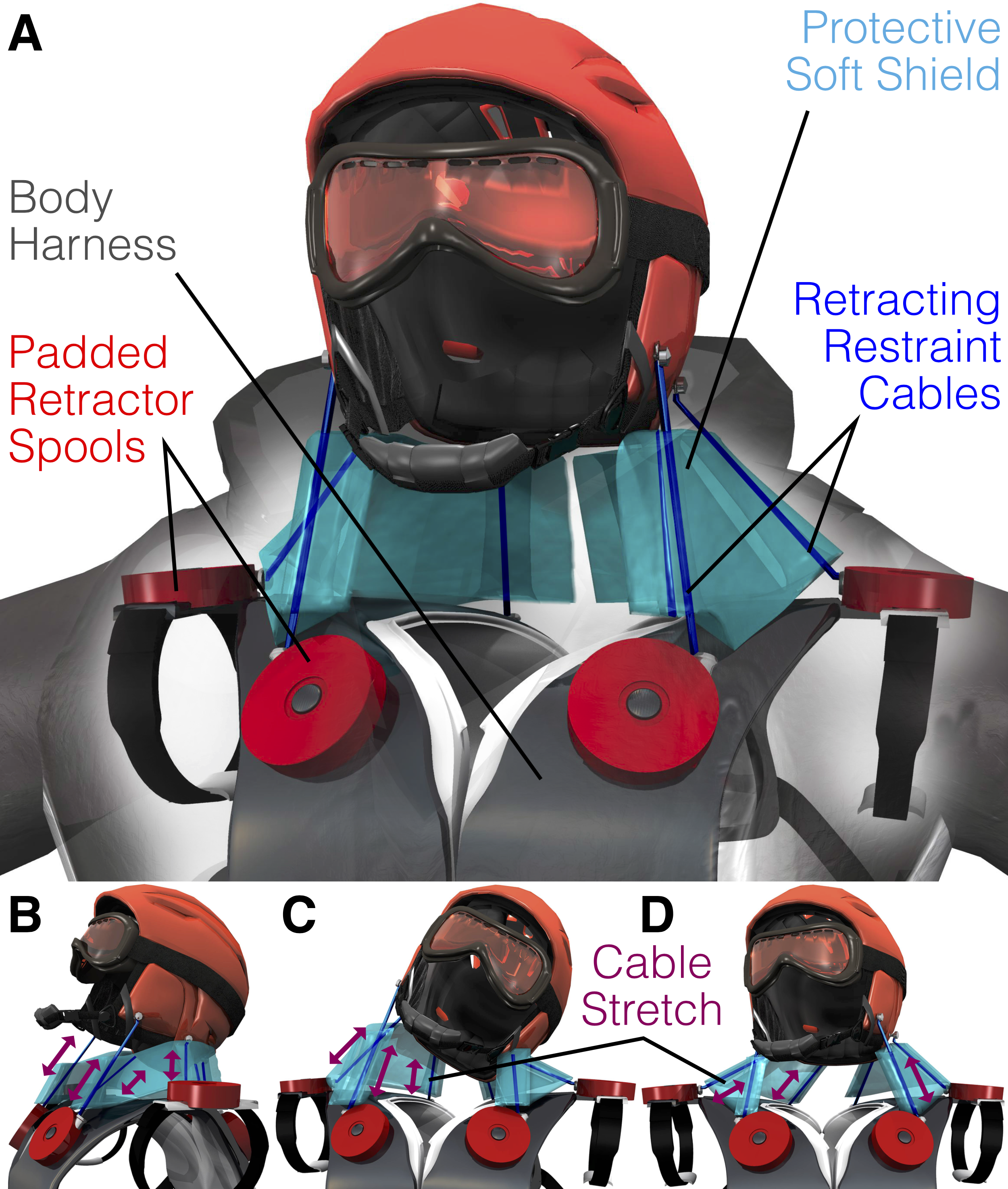
Summary: Brain injury occurs either due to (1) focal trauma from concentrated forces causing skull fracture/deformation that impinges on the brain, or (2) diffuse trauma from acceleration causing inertial loading deep within the brain. Focal brain trauma is largely a solved problem due to ubiquitous helmet use as concentrated forces are effectively distributed and therapeutics are on the horizon for meningeal injury. However, diffuse inertial injury is still highly prevalent despite helmet use. Whether helmeted or not, millions of people suffer from mTBI each year, which could be prevented if we address a basic problem: the human neck is the weak link in the chain. Helmets attempt to reduce the contact force on the head but run up against practical and theoretical limits, ultimately requiring the neck to do the critical work to prevent high inertial accelerations and resulting diffuse injury. Among primates, humans have the smallest neck/head area ratio, rendering the relatively weak human neck ineffective at restraining head acceleration during collision. When exposed to high loading inputs, the 5kg human head will remain suscep- tible to inertial injury until the neck is augmented with a restraint system that can provide safe operation while not impeding normal voluntary motion. We are convinced that a neck restraint can address the problem of inertial brain injury. Using an acceleration-triggered restraint system, we have invented an approach to selectively restrain head motion by transferring force to the torso. Similar to the action of neck muscles, tension in the restraint system results in compressive force on the cervical spine which must be maintained within safe levels (4.5kN based on reports by the National Highway Traffic Safety Administration (NHTSA). Our invention can feasibly prevent diffuse brain trauma by achieving three design goals: (1) reduce amplitude and duration of acceleration impulse, (2) permit voluntary movement, and (3) maintain safe cervical spine compression loads. |

|
Material characterization |

|
Summary: While advances in computational models of mechanical phenomena have made it possible to simulate dynamically complex problems in biomechanics, accurate material models for soft tissues, particularly brain tissue, have proven to be very challenging. We study the viscoelastic constitutive model of bovine brain tissue under various loading conditions. While many studies have investigated the mechanical behavior ofbrain tissue under various loading conditions, there has not been a clear explanation for variation reported for material properties ofbrain tissue. The current study compares the ex-vivo mechanical properties of brain tissue under two loading modes, namely compression and tension, and aims to explain the differences observed by closely examining the microstructure under loading. |